View Featured Offers >>
34608
PTMScan® HS K-ε-GG Remnant Magnetic Immunoaffinity Beads
Proteomic Analysis Products
Monoclonal Antibody
R
Recombinant

PTMScan® HS K-ε-GG Remnant Magnetic Immunoaffinity Beads #34608

Citations (0)
Motif analysis using all ubiquitin remnant peptides enriched and identified by PTMScan® HS K-ε-GG Remnant Magnetic Immunoaffinity Beads from three different samples. One milligram each of mouse brain, mouse liver, and MKN-45 human gastric cancer cells were independently digested with trypsin and immunoprecipitated with PTMScan® HS K-ε-GG Remnant Magnetic Immunoaffinity Beads. Orbitrap Fusion Lumos mass spectrometer analysis identified a total of 8,928 non-redundant sites. The motif logo shows that the K-ε-GG antibody is a general motif antibody that recognizes the K-ε-GG motif independent of protein context, without other amino acid preferences
To Purchase # 34608
Cat. # Size Qty. Price
34608S
200 µl

Supporting Data

REACTIVITY
SENSITIVITY Endogenous
MW (kDa)
Source/Isotype Rabbit IgG

Application Key:

  • WB-Western Blot
  • IP-Immunoprecipitation
  • IHC-Immunohistochemistry
  • ChIP-Chromatin Immunoprecipitation
  • C&R-CUT&RUN
  • C&T-CUT&Tag
  • DB-Dot Blot
  • eCLIP-eCLIP
  • IF-Immunofluorescence
  • F-Flow Cytometry

Species Cross-Reactivity Key:

  • H-Human
  • M-Mouse
  • R-Rat
  • Hm-Hamster
  • Mk-Monkey
  • Vir-Virus
  • Mi-Mink
  • C-Chicken
  • Dm-D. melanogaster
  • X-Xenopus
  • Z-Zebrafish
  • B-Bovine
  • Dg-Dog
  • Pg-Pig
  • Sc-S. cerevisiae
  • Ce-C. elegans
  • Hr-Horse
  • GP-Guinea Pig
  • Rab-Rabbit
  • All-All Species Expected

Product Description

PTMScan® HS is an enhanced PTMScan® methodology with improved identification of post-translationally modified peptides. PTMScan® technology employs a proprietary methodology from Cell Signaling Technology (CST) for peptide enrichment by immunoprecipitation using a specific bead-conjugated antibody in conjunction with liquid chromatography tandem mass spectrometry (LC-MS/MS) for quantitative profiling of post-translational modification (PTM) sites in cellular proteins. PTMs that can be analyzed by PTMScan® technology include phosphorylation, ubiquitination, acetylation, and methylation, among others. The technology enables researchers to isolate, identify, and quantitate large numbers of post-translationally modified cellular peptides with a high degree of specificity and sensitivity (HS), providing a global overview of PTMs in cell and tissue samples without bias about where the modified sites occur. For more information on PTMScan® products and services, please visit Proteomics Resource Center.

Storage

Antibody magnetic beads supplied in PBS buffer containing 0.1% Tween 20. Store at 4°C.

Protocol

PRINT

View >Collapse >

Day 1: Digest Proteins to Peptides
PTM Peptide IP

Materials & Reagents

Supplied Reagents:
  • PTMScan® HS Immunoaffinity Magnetic Beads
Additional Reagents (Not Supplied):
  • 200 mM HEPES, pH 8.0 (Cell Signaling Technology, #44686)
  • Phosphatase Inhibitor Cocktail (100X) (Cell Signaling Technology, #5870)
  • Urea, Ultrapure, PTMScan® Qualified (Cell Signaling Technology, #60055)
  • Iodoacetamide, PTMScan® Qualified (Cell Signaling Technology, #88931)
  • DTT (Dithiothreitol) (Cell Signaling Technology, #7016)
  • PTMScan® Trypsin, TPCK-Treated (Cell Signaling Technology, #56296)
  • PTMScan® Wild Type Alpha-Lytic Protease (WaLP) (Cell Signaling Technology, #33036)
  • 1 mM Hydrochloric acid (HCl)
  • Sep-Pak Vac 1 cc (50 mg) C18 Cartridges (Waters, #WAT054955)
  • Acetonitrile (Thermo Scientific, 51101)
  • Water, LC-MS Grade (Burdick and Jackson) (Cell Signaling Technology, #27732)
  • Trifluoroacetic acid (ThermoFisher Scientific, 28904)
  • 20X Phosphate Buffered Saline (Cell Signaling Technology, #9808)
  • BCA Protein Assay Kit (Cell Signaling Technology, #7780)
  • PTMScan® HS IAP Bind/Wash Buffer Kit (Cell Signaling Technology, #18494)
Equipment Not Included:
  • 1.5 mL microcentrifuge tubes
  • Magnetic rack for 1.5 mL microcentrifuge tubes (Cell Signaling Technology, #7017 or #14654)
  • End-over-end rotator
  • Centrifuge capable of handling 1.5 mL tubes
  • Vacuum concentrator (Speed-Vac)
  • pH indicator strips for pH 0-14
  • Pierce C18 Spin Tips (ThermoFisher Scientific, 84850)

I. Cell Lysis and Protein Digestion

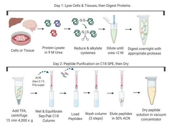
PTMScan® enrichment kits are compatible with many protein extraction, digestion, and purification protocols. Compatible workflows include in-solution digestion, those that use centrifugal reactors (FASP1, S-Trap cartridges2, or iST3), or magnetic bead precipitation (SP34). Regardless of the particular method selected, ensure that peptides are completely dry and free of lysis buffer components, lipids, and excess salts prior to using the immunoaffinity purification kit. Below is a general protocol that uses in-solution digestion followed by solid-phase extraction on Sep-Paks.

References:

  1. Wiśniewski, J. (2018) Methods Mol Biol 1841, 3-10. PMID: 30259475
  2. Zougman A. et.al. (2014) Proteomics 14(9), 1006-0. PMID: 24678027
  3. Kulak N.A. et.al. (2014) Nat Methods 11(3), 319-24. PMID: 24487582
  4. Hughes C.S. et. al. (2019) Nat Protocols 14(1), 68-85. PMID: 30464214

NOTE: Prepare solutions for cell lysis (Section I), C18 column purification (Section II), and peptide binding and washing during IAP enrichment (Section III) with reverse osmosis deionized (RODI) or equivalent grade water. Prepare solutions using HPLC grade water (Burdick and Jackson water) for the peptide elution (Section III) and the peptide concentration steps (Section IV).

A. Solutions and Reagents

    NOTE: Prepare solutions with RODI or equivalent grade water.

  1. Urea lysis buffer: 9 M urea, 20 mM HEPES pH 8.0, 2X Phosphatase Inhibitor Cocktail (Cell Signaling Technology, #5870). For 10 mL, weigh out 5.41 g of urea powder and dissolve it in 1 mL of 200 mM HEPES pH 8.0 stock and 4 mL RODI water. Mix well until the urea goes into solution completely. Add 200 µL of Phosphatase Inhibitor Cocktail (100X), and adjust the final urea lysis buffer volume to 10 mL total, such that the final concentration of phosphatase inhibitors is 2X.

    NOTE: The urea lysis buffer should be prepared fresh prior to each experiment. Do not include protease inhibitors.

    NOTE: Dissolving urea is an endothermic reaction. Urea lysis buffer preparation can be facilitated by placing a stir bar in the beaker and by using a warm (not hot) water bath on a stir plate. 9 M urea is used so that upon lysis, the final concentration is approximately 8 M. The urea lysis buffer should be used at room temperature. Placing the urea lysis buffer on ice will cause the urea to precipitate out of solution.

  2. DTT solution (1.25 M): Dissolve the 192.8 mg supplied in the product tube in 1.0 mL of RODI water.  Divide into 200 µL aliquots. Store at -20°C for up to one year. Thaw one aliquot for each experiment.
  3. Iodoacetamide solution: Dissolve 19 mg of iodoacetamide in water to a final volume of 1 mL. After weighing the powder, store in the dark and add water only immediately before use. The iodoacetamide solution should be prepared fresh prior to each experiment.
  4. Trypsin-TPCK: Store dry powder for up to two years at -80°C. Prepare 1 mg/mL stock in 1 mM HCl. Divide into 1 mL aliquots. Store aliquots at -80°C for up to one year.

B. Preparation of Cell or Tissue Lysate

Suspension Cells
  1. Grow approximately 1 x 10⁷ cells for each experimental condition or enough cells to produce approximately 1 mg of soluble protein. (If desired, scale up to 5 x 10⁷ cells.)
  2. Harvest cells by centrifugation at 130 x g for 5 min at room temperature. Carefully remove supernatant, wash cells with 20 mL of cold 1X PBS, centrifuge, remove PBS wash, and add 1 mL of urea lysis buffer (room temperature) to the cell pellet. Pipet the slurry up and down a few times (do not cool lysate on ice as this may cause precipitation of the urea).
Adherent Cells
  1. Grow approximately 1 x 10⁷ cells for each experimental condition or enough cells to produce approximately 1 mg of soluble protein. (If desired, scale up to 5 x 10⁷ cells.) The cell number corresponds to approximately one 150 mM culture dish (depending on the cell type), grown to 70-80% confluence.
  2. To harvest, remove media from the first dish by decanting, and let stand in a tilted position for 30 sec so the remaining medium flows to the bottom edge. Remove the remainder of the medium at the bottom edge with a P1000 micropipettor. Rinse each dish with 5 mL of cold PBS. Remove PBS as described above.
  3. Use 1 mL of lysis buffer per dish; e.g., start with 5 mL if five dishes will be harvested. Add 1 mL of urea lysis buffer (at room temperature) to the first dish, scrape the cells into the buffer, and let the dish stand in a tilted position after scraping the buffer to the bottom edge of the tilted dish.
  4. If multiple dishes will be combined to make a larger sample, remove the medium from the second dish as described above. Transfer the lysis buffer from the first dish to the second dish using a pipette, then tilt the first dish with the lid on for 30 sec and remove the remaining buffer from the dish and collect. Scrape cells from the second dish and repeat the process until the cells from all the dishes have been scraped into the lysis buffer. Collect all lysate in a 50 mL conical tube.

    NOTE: DO NOT place urea lysis buffer or culture dishes on ice during harvesting. Harvest cells using urea lysis buffer at room temperature. During lysis, the buffer becomes viscous due to DNA released from the cells.

Tissue Specimens
  1. Collect tissue specimens by flash freezing in liquid nitrogen. A specimen of approximately 20-50 mg (wet weight) should provide sufficient material for PTMScan® (1 mg of soluble protein or more).
  2. Place the still-frozen tissue in a 50 mL conical tube. Add a minimum of 2 mL of urea lysis buffer (use ~5 mL per 100 mg of wet tissue for larger specimens), or enough to submerge it completely.
  3. Lyse the tissue using a homogenizer. Wash the tool with RODI water in between each sample.

    NOTE: If desired, the PTMScan® protocol may be interrupted at this stage. The lysed cells or tissues can be frozen and stored at -80°C for several weeks.

C. Sonication and Centrifugation

  1. Using a microtip, sonicate lysate at 15 W output with 3 bursts of 15 sec each. Cool on ice for 1 min between each burst.
  2. Clear the lysate by centrifugation at 20,000 x g for 15 min at room temperature and transfer the protein extract (supernatant) into a new tube.

    NOTE: Centrifugation is performed at room temperature to prevent urea from precipitating out of solution.

    NOTE: Lysate sonication fragments DNA and reduces sample viscosity. Ensure that the sonicator tip is submerged in the lysate. If the sonicator tip is not submerged properly, it may induce foaming and degradation of your sample.

D. Protein Quantitation

  1. Measure soluble protein concentration using a colorimetric assay kit that is compatible with urea lysis buffer. The BCA assay is recommended.
  2. Normalize all samples so that equal amounts of protein are prepared for each condition and replicate. Alternatively, design the experiment so that equal numbers of cells are prepared for each sample. Add urea lysis buffer as needed to increase the volume of all samples to the same final amount.

E. Reduction and Alkylation of Proteins

  1. Add 1/278 volume of 1.25 M DTT to the cleared cell supernatant (e.g., 3.6 µL of 1.25 M DTT for 1 mL of protein extract), mix well, and incubate at room temperature for 60 min.
  2. Add 1/10 volume of iodoacetamide solution to the cleared cell supernatant (100 µL IAA for 1 mL lysate), mix well, and incubate for 15 min at room temperature in the dark.
  3. Dilute 200 mM HEPES pH 8.0 stock ten-fold with water to a final concentration of 20 mM HEPES pH 8.0.
  4. Dilute sample 4-fold with 20 mM HEPES, pH 8.0 to a final concentration of approximately 2 M urea, 20 mM HEPES pH 8.0. For example, add 3 mL 20 mM HEPES, pH 8.0 for 1 mL of lysate.

F. Protease Digestion

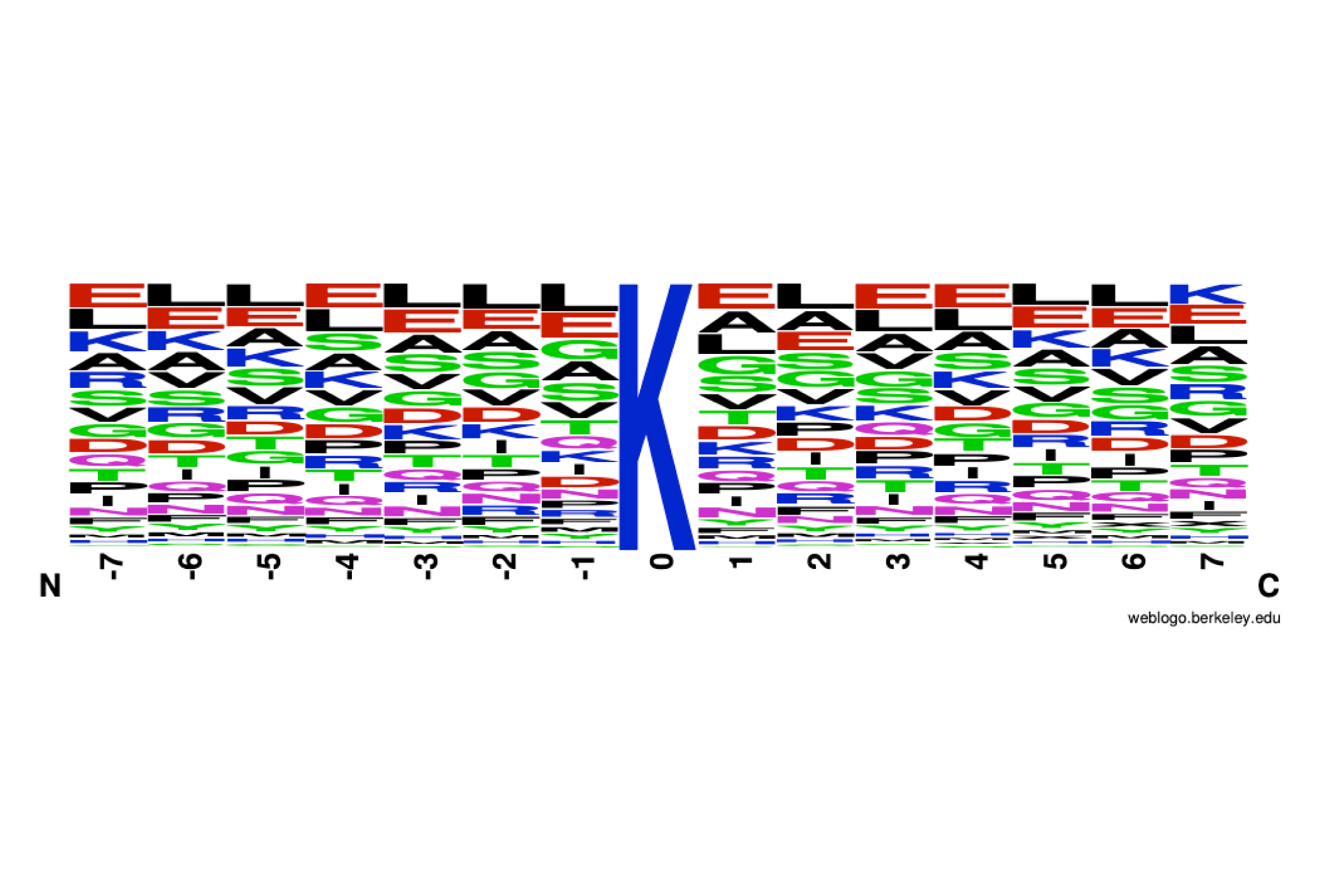
NOTE: Trypsin or WaLP is used for ubiquitinated or SUMOylated peptide analysis, respectively, in combination with the PTMScan® HS Ubiquitin/SUMO Remnant Motif (K-ε-GG) Kit (Cell Signaling Technology, #59322).

Trypsin Digestion
  1. Add Trypsin-TPCK at 1 mg : 37.5 mg (enzyme:substrate). This ratio requires 27 µL of 1 mg/mL PTMScan® Trypsin, TPCK-Treated (Cell Signaling Technology, #56296) stock for 1 mg lysate. Digest overnight at room temperature with mixing.
  2. Analyze the lysate before and after digest by SDS-PAGE to check for complete digestion.
WaLP Digestion

    NOTE: Use WaLP only for generating KGG remnants in SUMOylated protein experiments.

  1. Use 25 µL of WaLP stock (Cell Signaling Technology, #33036) for each 1 mg of protein lysate. Add WaLP at 1 mg : 100 mg (enzyme:substrate). This ratio requires 25 uL of 0.4 mg/mL WaLP (Cell Signaling Technology, #33036) stock for 1 mg lysate. Digest overnight at 37°C with mixing.
  2. Analyze the lysate before and after digest by SDS-PAGE to check for complete digestion.

II. C18 Purification of Lysate Peptides

NOTE: Purification of peptides is performed at room temperature on C18 reversed-phase Sep-Pak columns from Waters (#WAT054955).

NOTE: C18 purification uses reversed-phase (hydrophobic) solid-phase extraction. Peptides and lipids bind to the chromatographic material. Large molecules such as DNA, RNA, and most proteins, as well as hydrophilic molecules such as many small metabolites, are separated from peptides using this technique. Peptides are eluted from the column with 50% acetonitrile (ACN) and separated from lipids and proteins, which elute at approximately 60% ACN and above.

NOTE: About 2.5 mg of protease-digested peptides can be purified from one C18 column. Purify peptides immediately after proteolytic digestion.

A. Solutions and Reagents

    NOTE: Prepare solutions with LCMS grade reagents (e.g., water with Cell Signaling Technology, #27732; trifluoroacetic acid (TFA) with ThermoFisher Scientific, 28903; and acetonitrile (ACN) with Thermo Scientific, 51101). All percentage specifications for solutions are vol/vol.

  1. 20% trifluoroacetic acid (TFA): add 1 mL TFA to water to a total volume of 5 mL.
  2. Solvent A (0.1% TFA): add 1 mL of 20% TFA to 199 mL water.
  3. Solvent B (0.1% TFA, 50% acetonitrile): add 5 mL of acetonitrile (ACN) and 50 µL of 20% TFA to 4.95 mL of water.
  4. Wash buffer (0.1% TFA, 5% acetonitrile): For 10 mL of wash buffer, add 50 µL of 20% TFA to 9.45 mL of water, then add 500 µL of acetonitrile.

    NOTE: Organic solvents are volatile. Tubes containing small volumes of these solutions should be prepared immediately before use and should be kept capped as much as possible because the organic components evaporate quickly.

B. Acidification of Digested Cell Lysate

    NOTE: Before loading the peptides from the digested sample on the column, they must be acidified with TFA for efficient peptide binding. The acidification step helps remove fatty acids from the digested peptide mixture.

  1. Add 1/20 volume of 20% TFA to the digest for a final concentration of 1% TFA. Check the pH by spotting a small amount of peptide sample on a pH strip (the pH should be under 3). After acidification, allow precipitate to form by allowing the sample to stand for 15 min on ice.
  2. Centrifuge the acidified peptide solution for 15 min at 4,000 x g at room temperature to remove the precipitate. Transfer peptide-containing supernatant into a new 50 mL conical tube without dislodging the precipitated material.

C. Peptide Purification

    NOTE: Application of all solutions can be performed with a vacuum manifold or by gravity flow. If using vacuum, keep flow rates below approximately 0.33 mL/min for most steps. Sample loading should be done by gravity flow to maximize recovery.

  1. Pre-wet the column with 0.5 mL 100% ACN.
  2. Equilibrate the column with 1 mL of Solvent A (0.1% TFA). Repeat this step once.
  3. Load acidified and cleared digest (from Section B) one mL at a time until all of the sample has been loaded. Load sample by gravity flow, without applying vacuum.
  4. Wash with 1 mL of Solvent A (0.1% TFA). Repeat this step once.
  5. Wash with 0.5 mL of Wash buffer (5% ACN/0.1% TFA).
  6. Place columns above new 1.5 mL polypropylene tubes to collect eluate. Elute peptides with a sequential wash of 3 x 0.2 mL of Solvent B (0.1% TFA, 50% acetonitrile).
  7. Dry the peptide solution in a vacuum concentrator (Speed-Vac) set to ambient temperature overnight or until completely dry. The pellet should be visible at the end.

    NOTE: Peptide solutions may be frozen at -80°C for 1 hr or longer before placing in the Speed-Vac; this will prevent full tubes from spilling when placed at an angle to dry.

    NOTE: A standard lyophilization apparatus is also acceptable in place of a vacuum concentrator.

    NOTE: Dry, digested peptides are stable at -80°C for several months (seal the closed tube with parafilm for storage). The PTMScan® procedure can be interrupted before or after drying. Once the dry peptide is dissolved in 1X HS IAP Bind Buffer #1 (see next section), continue to the end of the procedure.

III. Immunoaffinity Purification (IAP)

A. Prepare solutions

  1. Thaw components of PTMScan® HS IAP Bind/Wash Buffer Kit #18494 at room temperature and return to ice once all contents are dissolved. If necessary, dissolve by shaking gently.
  2. IAP Elution Buffer: 0.15% TFA + 99.85% water. Add 11.3 µL of 20% TFA stock to 1.5 mL of LCMS grade water. Prepare this solution in a container that has never been exposed to soap, as detergents will interfere with LCMS analysis.

B. Procedure

  1. Centrifuge the dry peptides (approximately 1 mg is recommended for a PTMScan® HS experiment) for 5 min at 2,000 x g at room temperature to collect all material at the bottom of the tube. Add 1.5 mL of 1X HS IAP Bind Buffer #1 to dried peptides. Resuspend pellets mechanically by pipetting repeatedly, taking care not to introduce excessive bubbles into the solution.

    NOTE: Tubes can be shaken gently at room temperature using a vortexer or thermomixer for 5 min or placed in a sonicator bath for 2 min to ensure complete solubilization, if necessary.

    NOTE: After dissolving the peptide, check the pH of the peptide solution by spotting a small volume on pH indicator paper. The pH should be close to neutral (no lower than 7.0). If necessary, add 2 µL of 1 M Tris base at a time until the pH is at 7.0.

  2. Clear solution by centrifugation for 5 min at 10,000 x g at 4°C. Cool on ice.

    NOTE: There may be a small, insoluble pellet. Transfer supernatant to a clean tube and discard the pellet.

  3. Briefly spin the vial of antibody-bead slurry at no more than 2,000 x g for 2-5 sec to bring down any buffer and beads clinging to the sides and cap of the vial. Pipet the antibody-bead slurry gently to obtain a uniform suspension of beads, then take out 20 µL of bead slurry and place into a 1.5 mL microcentrifuge tube for each sample. Remix the bead stock before each pipetting step. Verify that each sample gets an equal aliquot of beads to ensure reproducible results.
  4. Transfer 1 mL of ice-cold 1X PBS into the 1.5 mL microcentrifuge tube, mix buffer with beads by inverting the tube five times. Place the tube on a magnetic separation rack. Wait 10 sec or until beads are attracted to the magnet. Carefully remove the PBS buffer. Repeat bead washing with 1 mL of 1X PBS three times for a total of FOUR PBS washes.
  5. Transfer the soluble peptide solution into the tube containing antibody beads and discard the insoluble pellet. Avoid creating bubbles upon pipetting.
  6. Tighten the cap and seal the top of the tube with plastic or a caplock clip to avoid leakage. Incubate on an end-over-end rotator for 2 hr at 4°C.

    NOTE: Ensure the beads remain in suspension while rotating and that bubbles do not collect at the bottom of the tube as this will prevent proper bead and sample mixing.

  7. Briefly spin the tube at no more than 2,000 x g for 2-5 sec to bring down the beads and solution clinging to the sides and cap. Place the tube in the magnetic separation rack for 10 sec. Transfer the unbound peptide solution to a microcentrifuge tube. Optional: Save this supernatant at -80°C for enrichments for other PTM types.

    NOTE: Keep the HS IAP Wash Buffer and LCMS Water on ice for the subsequent steps.

  8. Add 1 mL of chilled HS IAP Wash Buffer (1X) to the beads, mix by inverting the tube five times. Briefly centrifuge the tube, place the tube in the magnetic stand for 10 sec and remove all wash buffer. Repeat three more times for a total of FOUR washes with HS IAP Wash Buffer.
  9. Add 1 mL of chilled LCMS water to the beads, mix by inverting the tube five times. Briefly centrifuge the tube, place the tube in the magnetic stand for 10 sec and remove all water. Repeat one more time for a total of TWO water washes.
  10. Add 50 µL of IAP Elution Buffer (0.15% TFA) to the beads, use a mixer such as an Eppendorf ThermoMixer set at <500 rpm to keep beads in suspension at room temperature for 10 min. Do not mix so vigorously that beads are splashed to the sides and cap of the tube. If there is no mixer available, tap the bottom of the tube several times (do not vortex), and let stand at room temperature for 10 min, mixing gently every 2-3 min.
  11. Briefly centrifuge the tube and place it in the magnetic rack. Transfer eluted sample to a new microcentrifuge tube.
  12. Add another 50 µL of IAP Elution Buffer (0.15% TFA) to the beads to repeat the elution step. Combine both eluates in the same microcentrifuge tube. Verify that no beads are transferred to the collection tube by setting the elution on the magnet for a few sec.

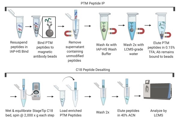
    NOTE: In this step, the post-translationally modified peptides of interest will be in the eluent.

IV. Concentration and Purification of Peptides for LCMS Analysis on C18 Tip

NOTE: We recognize there are many routine methods for concentrating peptides using commercial products, such as C18 tips (see below), that have been optimized for peptide desalting/concentration. Regardless of the particular method, we recommend that the method of choice be optimized for recovery and be amenable for peptide loading capacities of at least 10 µg.

C18 tips: Pierce C18 Spin Tips (ThermoFisher Scientific, 84850)

A. Solutions and Reagents

    NOTE: Prepare solutions with Burdick and Jackson water or other LCMS grade water. Organic solvents (trifluoroacetic acid, acetonitrile) should be of the highest grade.

    Recommended: Pierce Trifluoroacetic Acid (TFA), Sequencing grade (ThermoFisher Scientific, 28903) and Pierce Acetonitrile (ACN), LCMS Grade (ThermoFisher Scientific, 51101).

    Prepare all solutions in containers that have never been exposed to soap, as detergents will interfere with LCMS analysis.

  1. StageTip Wetting Solution (0.1% trifluoroacetic acid, 50% acetonitrile): add 50 µL of 20% trifluoroacetic acid to 4.95 mL LCMS water, then add 5 mL acetonitrile for 10 mL final volume.
  2. StageTip Equilibration & Washing Solution (0.1% trifluoroacetic acid): add 50 µL of 20% trifluoroacetic acid to 10 mL LCMS water.
  3. StageTip Elution Solution (0.1% trifluoroacetic acid, 40% acetonitrile): add 50 µL of 20% trifluoroacetic acid to 5.95 mL LCMS water, then add 4 mL acetonitrile for 10 mL final volume.

    NOTE: Organic solvents are volatile. Tubes containing small volumes of these solutions should be prepared immediately before use and should be kept capped as much as possible to prevent evaporation of organic components.

B. Procedure

    NOTE: All centrifugation steps in this section should be carried out at room temperature. Spin at 2,000 x g or a speed that passes all the solution through the tip in approximately 3 min.

  1. Cut the lid off of a 1.5 mL tube and place an adapter on top. Place a C18 tip in the adapter. There should be enough room in the tube to collect ~100 µL of liquid without touching the C18 material at the bottom of the tip.
  2. Equilibrate the C18 tip by pipetting 50 µL of Wetting Solution onto the top and centrifuging at 2,000 x g for approximately 3 min or until all the solution has passed through. Transfer the C18 tip to a clean collection tube.
  3. Add 50 µL of Equilibration & Washing Solution (0.1% TFA) and centrifuge 3 min. Repeat this step once for two total Equilibration & Washing steps.
  4. Load sample by passing IAP eluate through the C18 tip in two steps using 50 µL in each step.
  5. Wash the C18 tip by passing 50 µL of Equilibration & Washing Solution through two times.
  6. Elute peptides off the C18 tip by passing 10 µL of StageTip Elution Solution (40% ACN, 0.1% TFA) through two times. Pool the two resulting eluates.
  7. Dry down the C18 tip eluate from the C18 tip purification in a vacuum concentrator (Speed-Vac) and store the peptides dry at -20°C until LCMS analysis will begin. At that time, re-dissolve the peptides in an appropriate solvent for LCMS analysis, such as 5% acetonitrile, 0.1% TFA, 94.9% water (MS grade).

Protocol Id: 3148

Background

Ubiquitin is a conserved polypeptide unit that plays an important role in the ubiquitin-proteasome pathway. Ubiquitin can be covalently linked to many cellular proteins by the ubiquitination process, which targets proteins for degradation by the 26S proteasome. Three components are involved in the target protein-ubiquitin conjugation process. Ubiquitin is first activated by forming a thiolester complex with the activation component E1; the activated ubiquitin is subsequently transferred to the ubiquitin-carrier protein E2, then from E2 to ubiquitin ligase E3 for final delivery to the epsilon-NH2 of the target protein lysine residue (1-3). The ubiquitin-proteasome pathway has been implicated in a wide range of normal biological processes and in disease-related abnormalities. Several proteins such as IκB, p53, cdc25A, and Bcl-2 have been shown to be targets for the ubiquitin-proteasome process as part of regulation of cell cycle progression, differentiation, cell stress response, and apoptosis (4-7).

Small ubiquitin-related modifier 1, 2, and 3 (SUMO-1, -2, and -3) are members of the ubiquitin-like protein family (8). The covalent attachment of the SUMO-1, -2, or -3 (SUMOylation) to target proteins is analogous to ubiquitination.

Ubiquitin and the individual SUMO family members are all targeted to different proteins with diverse biological functions. Ubiquitin predominantly regulates degradation of its target (8). In contrast, SUMO-1 is conjugated to RanGAP, PML, p53, and IκB-α, regulates nuclear trafficking, forms subnuclear structures, and regulates transcriptional activity and protein stability (9-13). SUMO-2/-3 forms poly-(SUMO) chains, is conjugated to topoisomerase II and APP, regulates chromosomal segregation and cellular responses to environmental stress, and plays a role in the progression of Alzheimer's disease (14-17).

Limited Uses

Except as otherwise expressly agreed in a writing signed by a legally authorized representative of CST, the following terms apply to Products provided by CST, its affiliates or its distributors. Any Customer's terms and conditions that are in addition to, or different from, those contained herein, unless separately accepted in writing by a legally authorized representative of CST, are rejected and are of no force or effect.

Products are labeled with For Research Use Only or a similar labeling statement and have not been approved, cleared, or licensed by the FDA or other regulatory foreign or domestic entity, for any purpose. Customer shall not use any Product for any diagnostic or therapeutic purpose, or otherwise in any manner that conflicts with its labeling statement. Products sold or licensed by CST are provided for Customer as the end-user and solely for research and development uses. Any use of Product for diagnostic, prophylactic or therapeutic purposes, or any purchase of Product for resale (alone or as a component) or other commercial purpose, requires a separate license from CST. Customer shall (a) not sell, license, loan, donate or otherwise transfer or make available any Product to any third party, whether alone or in combination with other materials, or use the Products to manufacture any commercial products, (b) not copy, modify, reverse engineer, decompile, disassemble or otherwise attempt to discover the underlying structure or technology of the Products, or use the Products for the purpose of developing any products or services that would compete with CST products or services, (c) not alter or remove from the Products any trademarks, trade names, logos, patent or copyright notices or markings, (d) use the Products solely in accordance with CST Product Terms of Sale and any applicable documentation, and (e) comply with any license, terms of service or similar agreement with respect to any third party products or services used by Customer in connection with the Products.

For Research Use Only. Not for Use in Diagnostic Procedures.
Cell Signaling Technology is a trademark of Cell Signaling Technology, Inc.
All other trademarks are the property of their respective owners. Visit our Trademark Information page.