| Cat. # | Size | Qty. | Price |
|---|---|---|---|
| 26776T | 1 Kit (6 x 20 microliters) |
|
| Product Includes | Quantity | Applications | Reactivity | MW(kDa) | Isotype |
|---|---|---|---|---|---|
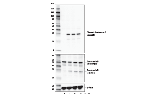
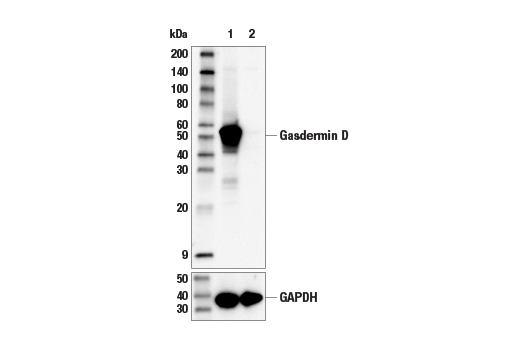
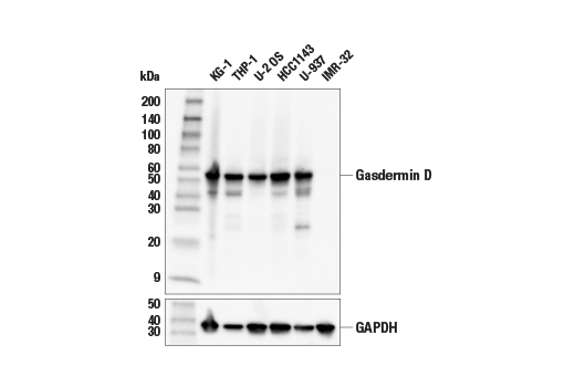
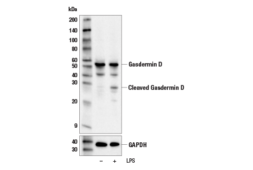
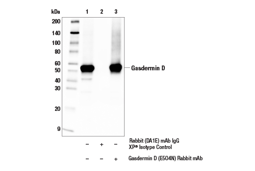
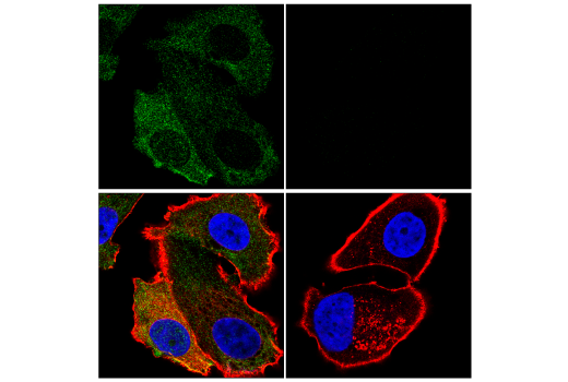
| Gasdermin D (E5O4N) Rabbit mAb 69469 | 20 µl |
|
H | 53, 30 | Rabbit IgG |
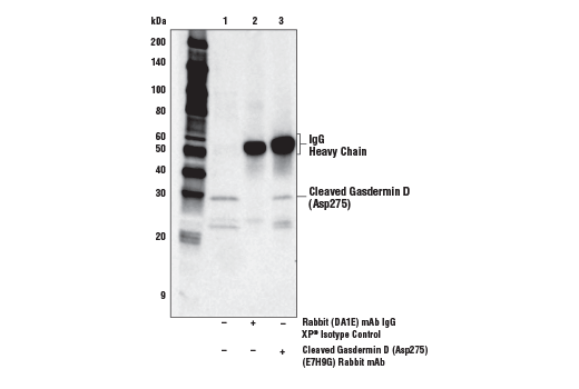
| Cleaved Gasdermin D (Asp275) (E7H9G) Rabbit mAb 36425 | 20 µl |
|
H | 30 | Rabbit IgG |
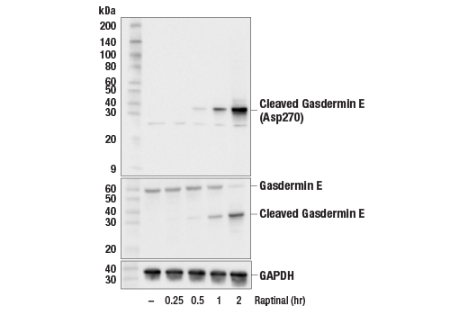
| Gasdermin E (E2X7E) Rabbit mAb 19453 | 20 µl |
|
H | 55, 30 | Rabbit IgG |
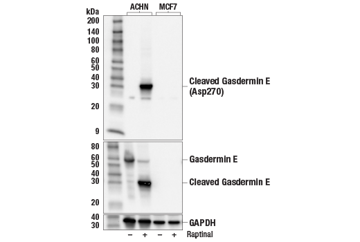
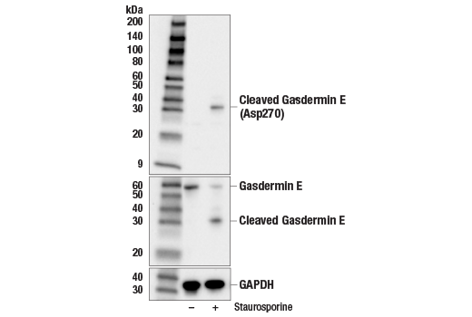
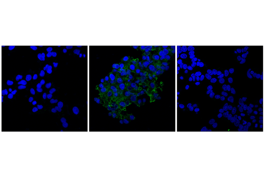
| Cleaved Gasdermin E (Asp270) (E8G4U) Rabbit mAb 55879 | 20 µl |
|
H | 30 | Rabbit IgG |
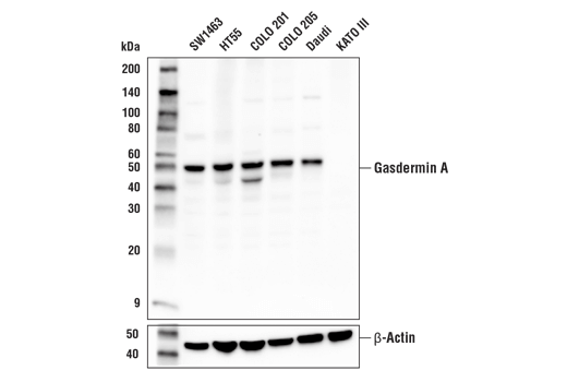
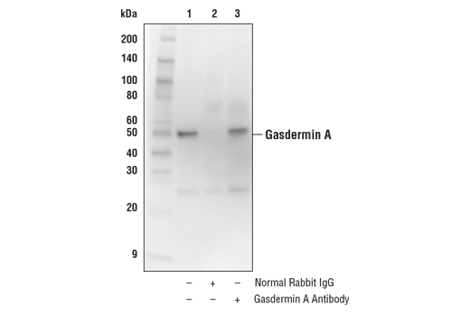
| Gasdermin A Antibody 49307 | 20 µl |
|
H | 49 | Rabbit |
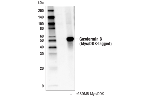
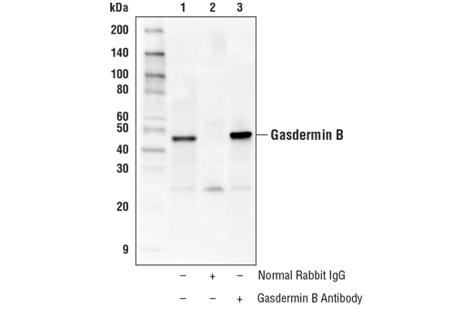
| Gasdermin B Antibody 76439 | 20 µl |
|
H | 47 | Rabbit |
| Anti-rabbit IgG, HRP-linked Antibody 7074 | 100 µl |
|
Goat |
Product Information
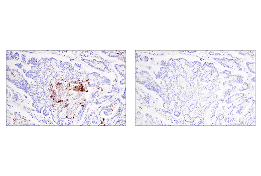
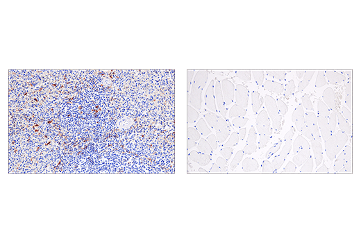
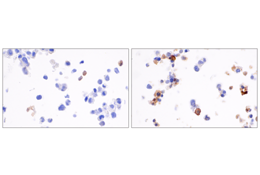
The gasdermin family, which includes GSDMA, GSDMB, GSDMC, GSDMD, and GSDME, has been shown to play a role in inflammation and cell death. Gasdermin D has been reported to have a critical role as a downstream effector of pyroptosis (1,2). Pyroptosis is a lytic type of cell death triggered by inflammasomes, multiprotein complexes assembled in response to pathogen-associated molecular patterns (PAMPs) or danger-associated molecular patterns (DAMPs) that result in the activation of caspase-1 and subsequent cleavage of pro-inflammatory cytokines IL-1β and IL-18 (3). Gasdermin D was identified by two independent groups as a substrate of inflammatory caspases, caspase-1 and caspase-11/4/5, producing two fragments: GSDMD-N and GSDMD-C. Cleavage results in release of an intramolecular inhibitory interaction between the N- and C-terminal domains, allowing the N-terminal fragment GSDMD-N to initiate pyroptosis through the formation of pores on the plasma membrane (4-7).
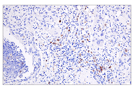
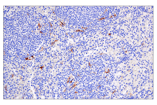
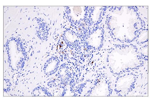
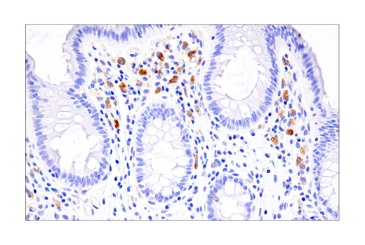
Like other gasdermin family members, Gasdermin E (also called DFNA5) contains an amino-terminal pore forming domain that triggers pyroptosis. Cleavage of Gasdermin E at Asp270 is induced by apoptotic-associated caspase-3, converting apoptotic signals to pyroptosis (8). In addition, cleavage of Gasdermin E can be induced by Granzyme B secreted by NK cells and contributes to tumor suppressive activity (9). Gasdermin E expression is suppressed in several types of cancer, including gastric, colorectal, and breast carcinoma, and may be associated with decreased survival (10-12). In contrast, an increase in Gasdermin E, including the amino-terminal pore-forming fragment, is associated with conditions of excessive inflammation (13-15). Gasdermin A (GSDMA) is preferentially expressed in the epithelium of the skin and gastrointestinal tract and is frequently suppressed in gastric cancer (16-18). Gasdermin B (GSDMB) has been reported to be upregulated in several tumor types, and in breast cancer has been associated with metastasis and poor prognosis (19,20). In addition, Gasdermin B has been associated with immune disorders, including asthma (21,22). Gasdermin B can be cleaved by Granzyme A secreted from cytotoxic lymphocytes leading to pyroptotic cell death (23).
Except as otherwise expressly agreed in a writing signed by a legally authorized representative of CST, the following terms apply to Products provided by CST, its affiliates or its distributors. Any Customer's terms and conditions that are in addition to, or different from, those contained herein, unless separately accepted in writing by a legally authorized representative of CST, are rejected and are of no force or effect.
Products are labeled with For Research Use Only or a similar labeling statement and have not been approved, cleared, or licensed by the FDA or other regulatory foreign or domestic entity, for any purpose. Customer shall not use any Product for any diagnostic or therapeutic purpose, or otherwise in any manner that conflicts with its labeling statement. Products sold or licensed by CST are provided for Customer as the end-user and solely for research and development uses. Any use of Product for diagnostic, prophylactic or therapeutic purposes, or any purchase of Product for resale (alone or as a component) or other commercial purpose, requires a separate license from CST. Customer shall (a) not sell, license, loan, donate or otherwise transfer or make available any Product to any third party, whether alone or in combination with other materials, or use the Products to manufacture any commercial products, (b) not copy, modify, reverse engineer, decompile, disassemble or otherwise attempt to discover the underlying structure or technology of the Products, or use the Products for the purpose of developing any products or services that would compete with CST products or services, (c) not alter or remove from the Products any trademarks, trade names, logos, patent or copyright notices or markings, (d) use the Products solely in accordance with CST Product Terms of Sale and any applicable documentation, and (e) comply with any license, terms of service or similar agreement with respect to any third party products or services used by Customer in connection with the Products.