Antibodies are only one of the factors responsible for experimental success. It is just as important to select companion products that help the antibody perform at its best. The companion products offered by CST are the same reagents our scientists use to validate our primary antibodies for immunohistochemistry (IHC). These products have been thoroughly optimized to work with our primary antibodies and protocols, so you can feel confident in your results. To browse the full offering of IHC companion products, click here.
The chemical crosslinks created during fixation can prevent antibody binding by inhibiting access to the antigen; therefore, it is necessary to reverse crosslinks using a procedure called antigen retrieval (also known as antigen unmasking or epitope retrieval). Antigen retrieval can be achieved through a heat-induced method (heat-induced epitope retrieval, or HIER) or through enzymatic digestion. During the antibody validation process, CST determines the optimal procedure for each primary antibody and includes the information on the product datasheet. SignalStain® Citrate Unmasking Solution (10X) #14746 and SignalStain® EDTA Unmasking Solution (10X) #14747 are two options offered by CST for antigen retrieval.
Normal Goat Serum #5425 is an ideal blocking reagent for chromogenic and fluorescent IHC applications. Tris Buffered Saline with Tween 20 (TBST-10X) #9997 and Animal-Free Blocking Solution (5X) #15019 are also available. Please consult the product-specific datasheet for the optimal buffers for your antibody of interest.
The primary antibody is a critical component of any IHC assay and has a direct effect upon data quality. A poor primary antibody can result in uninterpretable or misleading results. It takes more than a positive staining in a single tissue sample for an antibody to be approved for use in IHC. Antibodies from CST undergo a stringent validation procedure to ensure the antibody detects the target accurately. To browse our highly validated IHC-P antibodies, click here.
Antibody diluent can dramatically affect signal strength. SignalStain® Antibody Diluent #8112 yields superior staining when used as the diluent for recommended antibodies. Always consult the product-specific protocol developed for each primary antibody to determine the recommended diluent for your target of interest by searching IHC-P validated antibodies here.
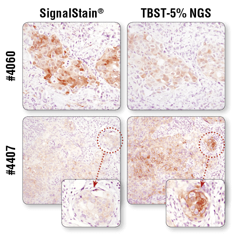
Antibody diluent can dramatically affect signal strength. IHC analysis of paraffin-embedded human breast carcinoma (top) and HCC827 xenograft (bottom) using Phospho-Akt (Ser473) (D9E) XP® Rabbit mAb #4060 or Phospho-EGF Receptor (Tyr1173) (53A5) Rabbit mAb #4407 after dilution in either SignalStain® Antibody Diluent (left) or TBST/5% NGS (right). As shown, a superior signal is achieved when #4060 is diluted in SignalStain® Antibody Diluent as compared with TBST/5% NGS. In contrast, #4407 performs better when diluted in TBST/5% NGS. Always check the product datasheet for the recommended diluent for your specific antibody.
You have applied your antibodies and obtained a signal, but is it a specific signal? Positive and negative controls instill confidence that your antibody is detecting its intended target. Prior to testing on tissue, antibody performance at CST is evaluated at the cellular level using a variety of cell lines and treatment conditions. For example, total protein specificity can be assessed through the use of positively and negatively expressing cell lines. Likewise, cells can be treated with biological or chemical modulators known to induce signaling changes to verify modification specificity, such as phosphorylation, acetylation, cleavage, etc. Phospho-specific antibodies can be further evaluated with phosphatase treatment. In addition, isotype control antibodies help rule out nonspecific staining of primary antibodies due to Fc receptor binding or other protein-protein interactions and should have the same immunoglobulin type as the test antibody. Click here to browse IHC control slides.
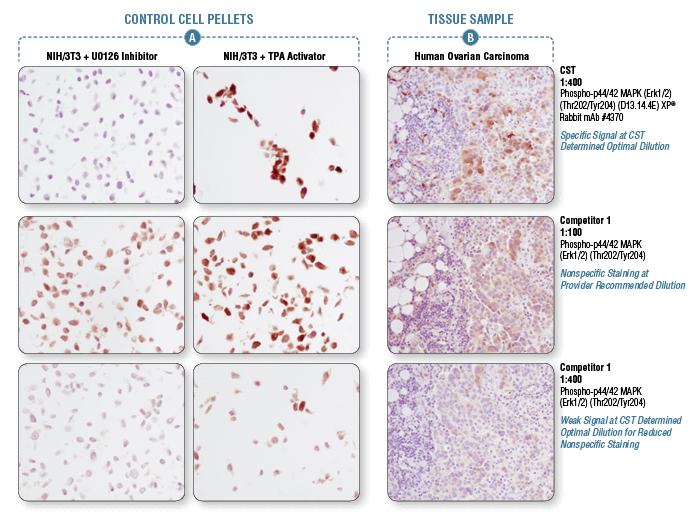
Appropriate controls are necessary to ensure specific signal. Phospho-p44/42 MAPK (Erk1/2) (Thr202/Tyr204) (D13.14.4E) XP® Rabbit mAb #4370 was compared to a competitor’s IHC-approved phospho-p44/42 (Tyr202/Tyr204) antibody. The optimal dilution of each antibody was individually evaluated to minimize nonspecific staining in NIH/3T3 cells (A) treated with the inhibitor U0126 (left) and to maximize specific signal in NIH/3T3 cells treated with the activator TPA #4174 (right). The determined optimal dilution for each antibody was utilized in IHC analysis of paraffin-embedded human ovarian carcinoma (B). Note that at the recommended dilution, the competitor’s antibody stains the inhibitor-treated cells (NIH/3T3 + U0126). At the CST determined optimal dilution, the competitor’s antibody no longer stains the inhibitor-treated cells, but also fails to stain tissue significantly. The CST antibody accurately stains both the cells and tissue.
CST scientists use Tris Buffered Saline with Tween 20 (TBST-10X) #9997 for ideal washing conditions in our IHC-P validated antibody protocols.
SignalStain® Boost IHC Detection Reagents #8125 and #8114 are highly sensitive, biotin-free, polymer-based reagents that avoid background staining caused by endogenous biotin. SignalStain® Boost IHC Detection Reagent (HRP, Mouse) #8125 and SignalStain® Boost IHC Detection reagent (HRP, Rabbit) #8114 can be used in IHC assays to detect mouse or rabbit primary antibodies, respectively.
Not all DAB substrate performs equally. SignalStain® DAB Substrate Kit #8059 offers high levels of sensitivity and works optimally with our primary antibodies.
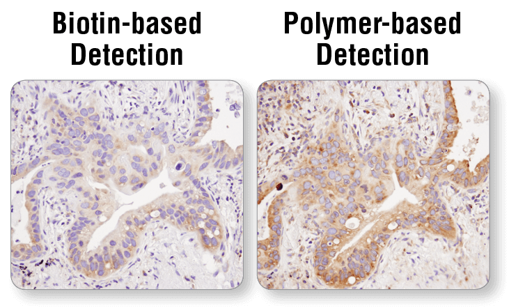
Polymer-based detection is more sensitive than biotin-based systems. IHC analysis of paraffin-embedded human lung carcinoma using S6 Ribosomal Protein (54D2) Mouse mAb #2317 and either biotin-based detection (left) or polymer-based detection (SignalStain® Boost IHC Detection Reagent #8125; right). As shown, polymer-based detection offers enhanced sensitivity and results in more robust staining.
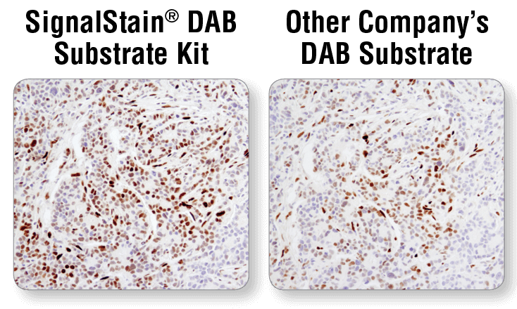
Not all DAB substrate performs equally. IHC analysis of paraffin-embedded human breast carcinoma using Phospho-Stat3 (Tyr705) (D3A7) XP® Rabbit mAb #9145. Chromogenic detection was performed using SignalStain® DAB Substrate Kit #8059 (upper), or DAB supplied by another company (lower). The SignalStain® DAB Substrate Kit produces a much stronger signal than the DAB supplied by another company.
Use SignalStain® Mounting Medium #14177 to ensure optimal conditions for slide mounting and storage.