Tau is a microtubule-associated protein expressed predominantly in the brain that functions to stabilize axonal microtubules. Tau exists as 8 different isoforms and can be phosphorylated at a number of unique sites, which, in turn, decreases its ability to bind microtubules. While Tau is phosphorylated in normal, healthy brain, it becomes hyperphosphorylated in neurodegenerative diseases including AD.
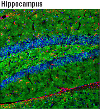
Confocal immunofluorescent analysis of mouse hippocampus using Tau (D1M9X) XP® Rabbit mAb (green) and GFAP (GA5) Mouse mAb #3670 (red). Blue pseudocolor = DRAQ5 #4084 (fluorescent DNA dye).
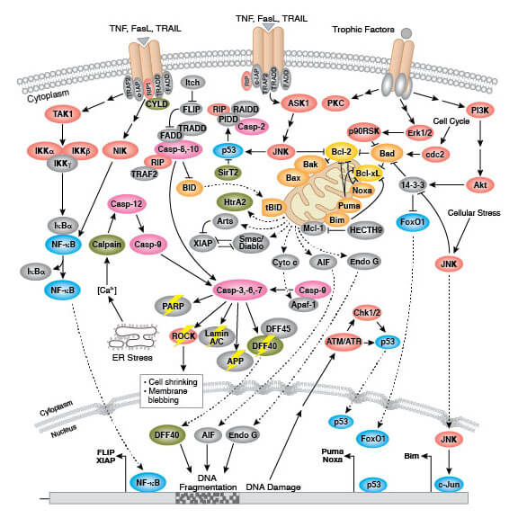
Neurofibrillary tangles found in Alzheimer’s disease result from the hyperphosphorylation of tau. Phosphorylation of tau can occur at many sites, typically by the kinases GSK3, CDK5, and results in a decreased ability to bind microtubules.
Hyperphosphorylation results in tau dissociation and subsequent destabilization of the microtubules and oligomerization of tau. Neurofibrillary tangles, as observed in Alzheimer’s disease and other tauopathies, are collections of paired helical filaments composed of hyperphosphorylated tau. It is thought that tau proteins are able to form prion-like “seeds” by which it can spread from neuron to neuron, and the presence of neurofibrillary tangles eventually leads to neuronal apoptosis and further pathogenesis of disease.
The levels of total and phosphorylated tau can be used to assist in the diagnosis and treatment of neurodegeneration. For example, total and phosphorylated tau are highly sensitive biomarkers that can predict the progression of mild cognitive impairment to Alzheimer’s disease.
The phosphorylation status of tau dramatically affects the intracellular distribution of the protein. Phosphorylated tau has been found in the neuronal soma of hippocampal neurons as well as immortalized GnRH neurons. Further evidence demonstrates that tau phosphorylation at Ser404 destabilizes microtubules as well as a link between Alzheimer’s disease and hyperphosphorylation at Ser404.
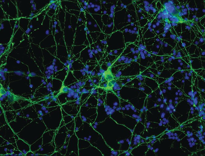
Confocal immunofluorescent analysis of mouse primary neurons using Phospho-Tau (Ser404) (D2Z4G) Rabbit mAb (IF preferred) (green). Blue pseudocolor = Hoescht 33342 #4082 (fluorescent DNA dye).
Monoclonal antibodies to tau are designed to recognize endogenous levels of tau protein as well as various forms of phosphorylated tau including Thr181, Ser199, Ser202, Thr205, Ser214, Thr231, Ser396, Ser404, and Ser416. The levels of tau aggregated in neurofibrillary tangles can be determined in a tissue sample through western blotting, immunoprecipitation, immunofluorescence, and immunohistochemistry.
In support of neurodegeneration research, a number of antibodies have been developed by scientists at Cell Signaling Technology that enable the analysis of tau and phosphorylated tau.