Mature immune cells differentiate from hematopoietic stem cells, that are found in the bone marrow, peripheral blood, and placenta. These hematopoietic stem cells can differentiate into the common myeloid progenitor (CMP) cell or the common lymphoid progenitor (CLP) cell. CMPs and CLPs give rise to the various cellular components of the innate and adaptive immune systems.
Groups of antibodies with similar binding patterns to leukocyte cell surface molecules at various stages of differentiation are used to identify “cluster of differentiation” (CD) antigens. These patterns of CD expression identify individual classes of immune cells and can be used to detect the presence of a specific immune cell in a heterogeneous population. For example, T Cells are characterized by the expression of CD3 but can be further subtyped as cytotoxic T Cells by the presence of CD8 or T helper cells by the presence of CD4.
Immunohistochemical immunophenotyping allows evaluation of the expression and tissue architecture of some proteins, which may not be possible by flow cytometric immunophenotyping. In addition, IHC observations may provide additional information regarding patterns of involvement and the cytological features of cells that cannot be obtained by flow cytometry.
CST has created a helpful table of common immune cell phenotyping markers, complete with links to additional information and related product information.
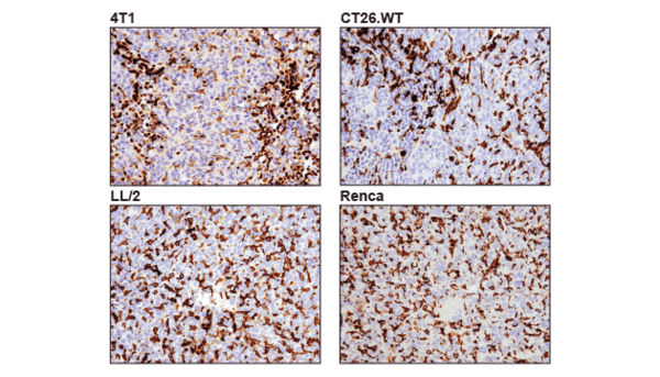
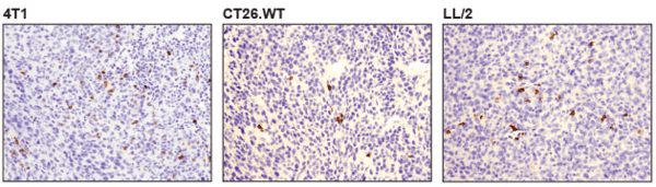
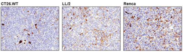
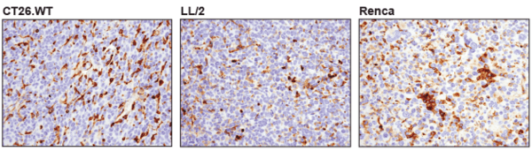
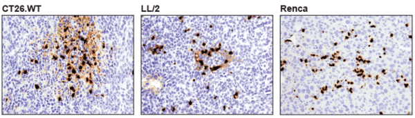
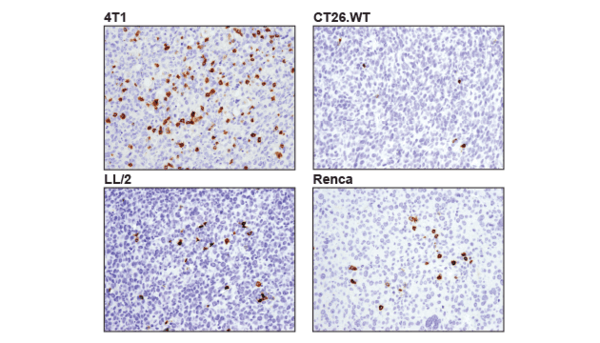
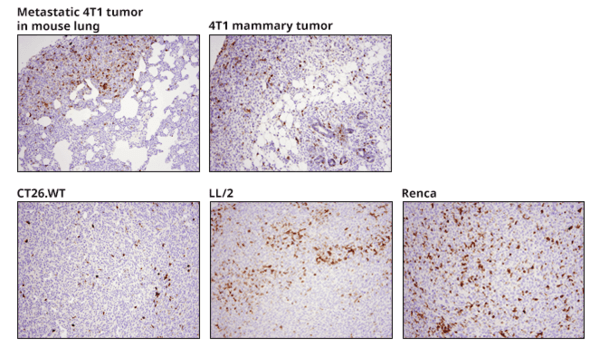
Mouse models are valuable pre-clinical research tools that are genetically and biologically similar to humans. However, mouse monoclonal antibodies suffer from secondary antibody-related background when used on mouse tissue. Rabbit monoclonal antibodies are an ideal solution to this problem. CST provides a constantly expanding portfolio of mouse-reactive rabbit monoclonal antibodies that demonstrate the exceptional specificity, sensitivity and performance required for credible results in IHC immunophenotyping. CST maintains high quality standards by using syngeneic mouse models, as well as normal mouse tissues, to confirm specificity in the same pre-clinical models our customers are using.
Flow Cytometry has some advantages over IHC. In particular, flow cytometry allows for: